![Diagnostic Imaging Knowledge Service [Diagnostic Imaging Tutor]](../imgs/img_diagnosis_01.jpg)
- Diagnostic Imaging Navigator [Doc.navi]
- Diagnostic Imaging Simulator [simu.Doc]
![Diagnostic Imaging Navigator [Doc.navi]](../imgs/img_diagnosis_02.png)
Doc.navi supports users to improve their skills for diagnosing
highly specialized modern medical imaging. In addition,
you’ll find both more efficient clinical management and
patients’ satisfaction through our Doc.navi support.
- More accurate
diagnosis by
listing possible diseases - Easy to take following
actions by presenting
the possible diseases - More efficient diagnosis
by retrieving and
showing the suspected
cases instantly
![Diagnostic Imaging Navigator [Doc.navi]](../imgs/img_diagnosis_03.jpg)
 Diagnostic imaging navigation
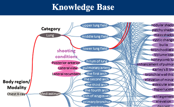
Doc.navi guides users to select very likely diagnosis step by step from “procedure”, “region”, and “basic finding”.
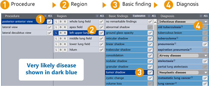
 Imaging cases search
Selected and useful case images have information of “patient attribute”, “definitive diagnosis”, “medical record” and “specialist’s explanation.” From “basic finding”, “diagnosis”, etc. imaging cases to compare your patient’s one with are searched immediately, which helps you to diagnose efficiently.
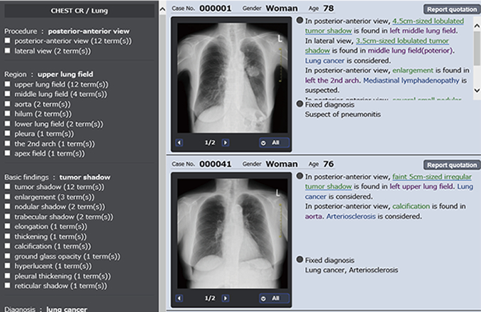
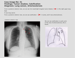
Case explanation
Each case imaging contains specific explanations commented by specialists
how they figured it out.

-

Support
filling medical recordMedical records are automatically and immediately prepared by selecting “ region”, “basic finding” and “diagnosis”. This function shortens the time for recording and your paper work.
-

Commentary
on findingsEasy to diagnose by reviewing the medical imagings that have pathognomonic findings.
-

Disease explanation
Simple explanations regarding “diagnostic result”, “explanation about disease”, “treatment plan”, “precautions”, etc. are displayed that can be printed for the patients.

Hyogo Prefectural Kakogawa Medical Center
Chief of Radiation Department and Director
(Palliative Medicine)
Eirou Sakai, M.D.,Ph.D.
Unsatisfied quality of medical imaging and wrong interpretation of the medical imaging are quite harmful for the patients. The quality of diagnosis of interns and residents and supportive reading by radiological technologists depend on the individuals. I do expect that Doc.navi will support users to improve their comprehensive capacity to read and interprete the medical imaging.
- This is a real professional tool for accurate diagnosis and supports making prompt decisions for the patients.
- When interpretation of medical imagings is fairly difficult, sample cases displayed guide us to make right diagnosis.
- Easy and supportive to explain diagnostic results to patients by showing patient' images.
- Easy and quick to prepare the medical records.
- Easy to understand the concept and procedure of diagnostic imaging.
- Supportive to restudy and recollect about the clinical findings learned while interns and residents as post graduate trainings.
![Diagnostic Imaging Navigator [simu.Doc]](../imgs/img_diagnosis_s01.jpg)
Build up your skill in pinpointing abnormalities and
interpreting symptoms from medical imaging by simulating
the diagnostic know-how of medical specialists and supervisors.
- Educate yourself online
-anytime and anywhere! - The most effective
cases selected from
leading university
hospitals in Japan - Build up your skill in
pointing out abnormalities
and interpreting symptoms
from medical imaging
 You can repeatedly drill yourself on the basics through practice questions.
A self-assessment function gives your performance levels.
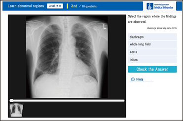
Typical exercises by the difficulty level
- Select any difficulty level of the contents to learn.
- Select an answer from 4 or 5 choices. Hints may help you.
- Check answers and explanations by "Check the Answer."
- Check your record and rank after answering all questions.
- You can try the exercise again by just clicking "Again."
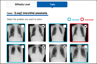
Specific exercises by the topics
- Select "Findings" and "Cases" to learn.
- Select an answer from 4 or 5 choices. Hints may help you.
- Check answers and explanations by "Check the Answer."
- Answer all questions, then check your records and performance.
Advice
of
"Basic"
- Check your performance and
the correct answers right
after finishing all questions. - Check your level and achievement
from your record list. - You can repeatedly learn
on specific cases.
 Clinical diagnostic imaging is shown on the screen.
Simulating diagnostic experiences improves your image interpretative skill.
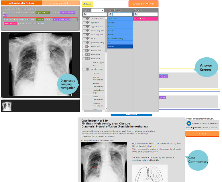
Exercise flow
- Set the scope of questions, difficulty level, and other items on the initial screen.
- Check case images and select terms from the Dianostic Imaging Navigaton.
- Compare your answers with correct answers and explanations.
- Answer all questions and then check "Record" to review your performance.
Advice
of
"Practice"
- Set a degree of difficulty
that matches
your own level. - By narrowing down answers using
Diagnostic Imaging Navigation
you can acquire the necessary thinking
for diagnostic process - After checking your answer,
deepen your understanding by
viewing tha case commentary
Recommended for from the beginners to the advanced learners.
The effective tool to improve the skill of each learner

- point 1
- Possible to select the contents for your skill
- You can select the contents from “Region”, “Finding” and “Diagnosis.” Set your learning preferences to match the skill you need.
- point 2
- 4 levels questions
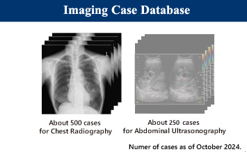
- About 1000 well-selected educational cases are presented as questions. They are divided into 4 levels, introduction, beginner, intermediate and advanced. You can set up questions according to your level.
- point 3
- Gradual improvement
- If you use the function “Hint” well, your level can up gradually. Get the most out of “Hint” when you just start learning. Improving your skill, you’ll reduce the use of “Hint” and step up your level gradually.
Users’ voice
The almost actual-size images
- It is good for me to observe clear case images well on the monitor. In addition, we can see not only the images with lesions but also the normal cases. This helped me a lot.

Simulation of reading images
- I simulated the know-how of interpretations such as where and how the lesions were and what they were suspected to be finally. This was good chance for me to think about images neccesary for radiological technologists.
Preparation before getting a position
- Knowing images necessary for diagnosis lets us get conscious of reading images useful for diagnosis and treatment. I want to acquire the skill and knowledge at clinical scenes.
![Diagnostic Imaging Navigator [Doc.navi]](../imgs/tab1_navi2_on.png)
![Diagnostic Imaging Simulator [simu.Doc]](../imgs/tab1_simu2_on.png)


Diagnostic Radiologist & Director
Naoki Mihara,
M.D.,Ph.D.
Former Director of Medical
Information Department,
Hiroshima University Hospital
Master the knack of picking out abnormal signs in medical imaging on sight!
The role of medical imaging are getting more and more important in today’s medical science. Development of the modern medical imaging technologies has been providing rather detailed and accurate images that are essential as one of the most important diagnostic tools. Medical images are not just simple data but are valuable information of patients. Interpretation is the work to change mere data of the images into valued information of the patients. Acquiring the skill to interpret the information from the medical imaging is not only the power to understand the patients but also contribute to medical science. Due to cost efficiency, chest radiographs are the most applicable examinations for diagnosing the lung diseases in the most clinics and hospitals. Chest radiographs contain a variety of symptoms, and often provide the suspected diseases. I recommend the people studying medicine to become specialists to interpret the medical imaging by systematic learning methods. Simu.Doc contains many different types of cases that train you to find abnormalities in the medical images at sight. I am proud to introduce our “simu.Doc” to help you as the most powerful tool to get the skills.